Overview
Bone age assessment (BAA) is a clinical procedure frequently performed in pediatric radiology to evaluate the stage of skeletal maturation based on a left hand and wrist radiograph. The determination of bone age plays an important role in diagnostic and therapeutic investigations of endocrine problems and growth disorders of children. The most commonly used method in the United States is the Greulich and Pyle (G&P) Hand Atlas, unchanged from its initial publication in 1950s. In addition, it was exclusively based on the Caucasian population and may not be fully applicable to the other racial descendents of today.
During the past ten years, we have developed a digital hand atlas (DHA) with 1,400 hand images of normal children, male and female from Asian, African American, Caucasian and Hispanic descent collected at the Children’s Hospital Los Angeles (CHLA) funded by NIH; and a fully automatic and robust computer-aided-diagnosis (CAD) method adapted to these four populations and two genders. With DHA and CAD method in the clinical environment as a useful tool, the future BAA workflow is shown in Figure 1.
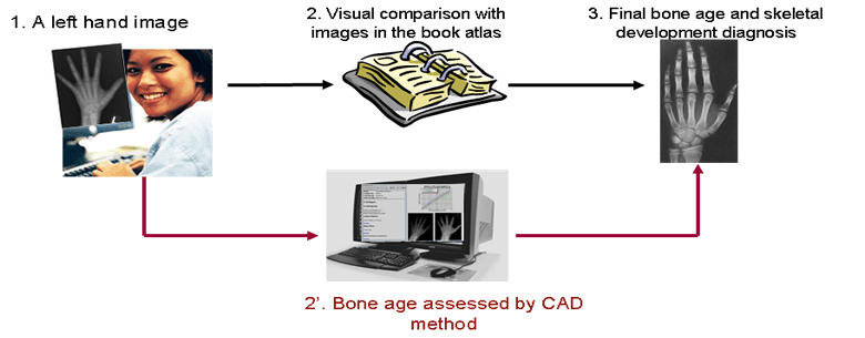
Figure 1. The ideal scenario of bone age assessment.
Materials and Methods
A total of 1,400 left hand and wrist radiographs of normal children, along with patients’ demographic data and radiologists’ readings were collected from Childrens Hospital Los Angeles, distributed in 19 groups (newborn, 1 – 18) of eight categories (i.e. Caucasian, African-American, Hispanic, and Asian), both male and female. A computer-aided-diagnosis method has been developed to determine the bone age based on two regions: phalangeal and carpal bone regions from each image. The integration of the third region: wrist joint is under development. (Figure 2)
Figure 2. An example left hand and wrist radiograph with super-imposed regions of interest.
Collaborating with Dr. Linda Vachon, chief of pediatric radiology at Los Angeles County Women’s and Children’s Hospital (LAC WCH), we are in implementation of the CAD system in clinical daily practice. Referring to Figure 3, the current clinical workflow of the BAA at LAC WCH is as follows: 1) Hand X-ray from the CR (Computed Radiography) modality is sent to PACS workstation (WS) where radiologists review images and perform BAA (Figure 3 upper dotted box). In the clinical implementation of the BAA CAD system, the additional workflow is as follows: 2) A second copy of image from radiology department is sent to the CAD server located at IPILAB; 3) The CAD BAA report will be available in 2-3 minutes for radiologists at LAC WCH to review; 4) The radiologist who reviewed this case will perform a second assessment which is captured and will be sent back to the server for tabulation and statistical analysis.
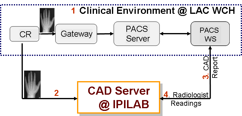
Figure 3. Workflow of CAD implementation at LAC WCH.
Clinical relevance/application:
The success of the clinical validation will lead to a revolutionary, innovative and cost-effective CAD method for real-time automatic BAA to assist radiologists.
Links (For Research and Education Purpose Only):
1) Web Image Library:
Download Option 1: Digital Hand Atlas http://ipilabmysql.usc.edu/newindex.php
Download Option 2: Download the entire Digital Hand Atlas (7GB) here
2) Plots of Chronological age, readings and CAD results: BAA graphs
3) Technical Details of Bone Age Assessment CAD:https://ipilab.usc.edu/research/content-php/
Publications
- Pietka E, Gertych A, Pospiech S, Cao F, Huang HK, Gilsanz V, “Computer Assisted Bone Age Assessment: Image Processing and Epiphyseal/Metaphyseal ROI Extraction,” IEEE Trans. Medical Imaging, 20, 715-729, 2001.
- Certification of Merit Award, Pietka E, Gertych A, Witko K, Remotely accessible computer Assisted skeletal Maturity Assessment, Proceedings of RSNA Conference, pp. 807, Chicago, 2004.
- Huang HK, Zhang A, Liu BJ, Zhou Z, Documet J, King N, Chan LWC, “Data Grid for Large-Scale Medical Image Archive and Analysis”, Proceedings of the 13th ACM International Conference on Multimedia, 2005
- Zhang A, Huang HK, Gertych A, Liu BJ, “Fuzzy System Design for Bone Age Assessment of Children in Multiple Regions of the Hand”, Scientific Poster (accepted), RSNA 2007
- Zhang A, Gertych A, Liu BJ, “Automatic Bone Age Assessment for Young Children from Newborn to 7-Year-Old Using Carpal Bones”, Computerized Medical Imaging and Graphics, May 2007
- Gertych A, Zhang A, Sayre, J, Pospiech-Kurkowska S, Huang HK, “Bone Age Assessment of Children using a Digital Hand Atlas,” Computerized Medical Imaging and Graphics, May, 2007.
- Zhang A, Sayre JW, Tsao S, Huang HK, Vachon L, “Cross-Racial Discrepancies of Growth Patterns in Bone Age Assessment”, Scientific Paper (accepted), RSNA 2007
- Tsao S, …wrist joint analysis [RSNA2007]
Contact: ipilab2013@gmail.com